The Kidney
The Kidney
The Kidney
Organs of the urinary system table
Organ | Explanation |
|---|---|
Kidney | Two bean-shaped organs that filter the blood |
Ureter | The tube that connects the kidney to the bladder |
Bladder | The organ that stores urine (excess water, salts and urea) after the kidney produces it |
Urethra | The tube that connects the bladder to the exterior; urine is released this way |
The human urinary system diagram
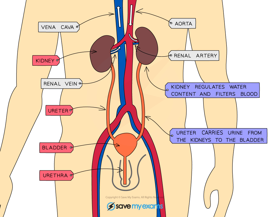
The urinary system in humans contains the kidneys and bladder, as well as the ureter and urethra
Kidney Structure
Kidney Structure
The Nephron
Each kidney contains around a million tiny structures called nephrons, also known as kidney tubules or renal tubules
The nephrons start in the cortex of the kidney, loop down into the medulla and back up to the cortex
The contents of the nephrons drain into the innermost part of the kidney and the urine collects there before it flows into the ureter to be carried to the bladder for storage
Nephron diagram
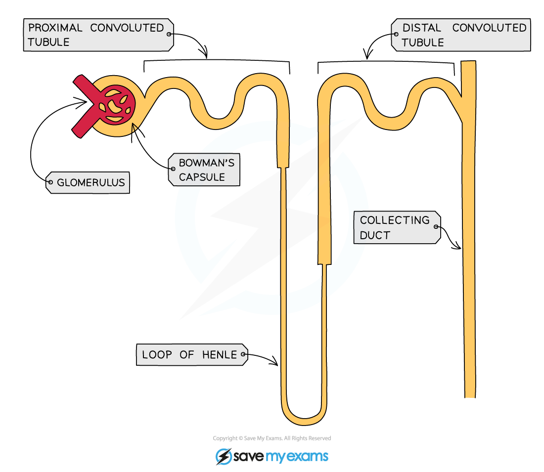
The kidney contains many tiny structures known as nephrons
Arterioles branch off the renal artery and lead to each nephron, where they form a knot of capillaries known as the glomerulus, which sits inside the cup-shaped Bowman’s capsule
The smaller molecules carried in the blood are forced out of the capillaries and into the Bowman’s capsule, where they form what is known as the filtrate
The substances forced out of the capillaries are
Glucose
Water
Urea
Salts
Some of these filtered molecules are useful and will be reabsorbed back into the blood further along the nephron
The urea, water and mineral salts that remain in the filtrate will form urine which is stored in the bladder
Filtration of blood diagram
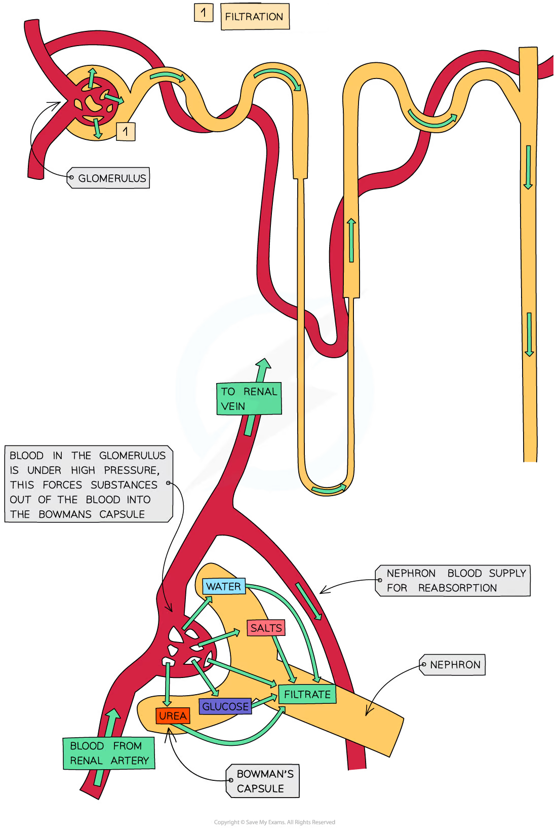
The blood is filtered by the glomerulus
Components of filtrate table
Component | Location of reabsorption |
|---|---|
Water | Loop of Henle and collecting duct |
Salts | Loop of Henle |
Glucose | Proximal convoluted tubule |
Urea | Not reabsorbed |
After the Bowman’s Capsule, glucose is the first substance to be reabsorbed; this occurs in the proximal convoluted tubule
As the filtrate passes through the Loop of Henle, necessary salts are also reabsorbed back into the blood
When salts are reabsorbed back into the blood, water follows by osmosis
Water is also reabsorbed from the collecting duct in different volumes, depending on the requirements of the body
Reabsorption diagram
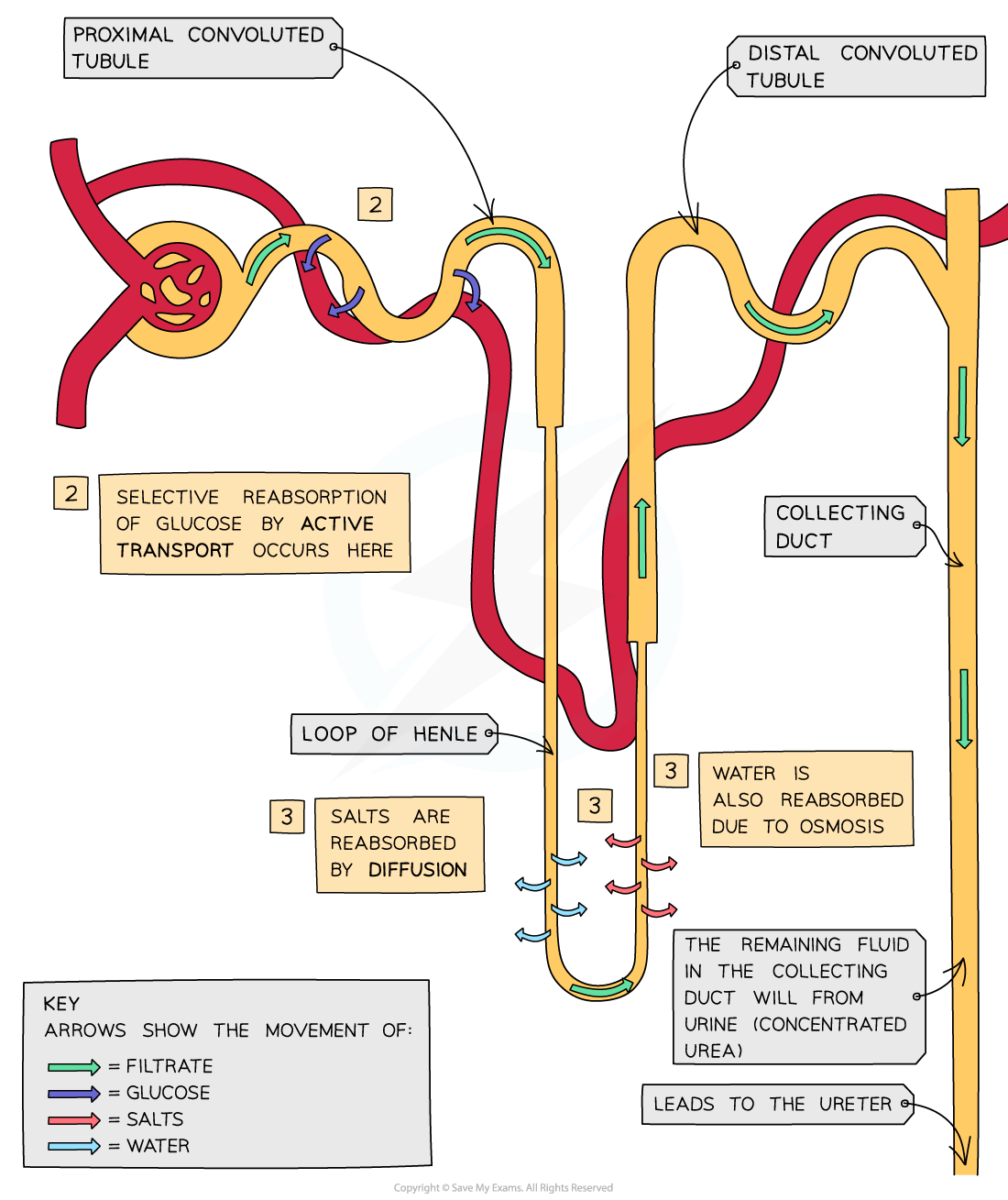
Reabsorption occurs primarily in the proximal convoluted tubule and loop of Henle