Animal & Plant Cells Under the Microscope
Examining Cells Under the Microscope
Examining Cells Under the Microscope
Viewing cells under the microscope
Many biological structures are too small to be seen by the naked eye
Optical microscopes are an invaluable tool for scientists as they allow for tissues, cells and larger organelles to be seen and studied
Light is directed through a thin layer of biological material that is supported on a glass slide
This light is focused through several lenses so that an image is visible through the eyepiece
Getting a visible image requires a very thin sample of biological tissue because light has to pass through the sample and into the lenses of the microscope
The most common specimens to observe under a light microscope are cheek cells (animal cells) and onion cells (plant cells)
A stain is often used to ensure cell structures are clearly visible under the microscope
General method
Specimens must be prepared on a microscope slide to be observed under a light microscope
This must be done carefully to avoid damaging the biological specimen and the structures within it
Preparing a slide using a liquid specimen:
Add a few drops of the sample to the slide using a pipette
Cover the liquid/smear with a coverslip and gently press down to remove air bubbles
Wear gloves to ensure there is no cross-contamination of foreign cells
Preparing a slide using a solid specimen:
Use scissors to cut a small sample of the tissue
Peel away or cut a very thin layer of cells from the tissue sample to be placed on the slide (using a scalpel or forceps)
Some tissue samples need to be treated with chemicals to kill/make the tissue rigid
Gently place a coverslip on top and press down to remove any air bubbles
A stain may be required to make the structures visible depending on the type of tissue being examined
Commonly used stains include methylene blue to stain cheek cells and iodine to stain onion cells
Take care when using sharp objects and wear gloves to prevent the stain from dying your skin
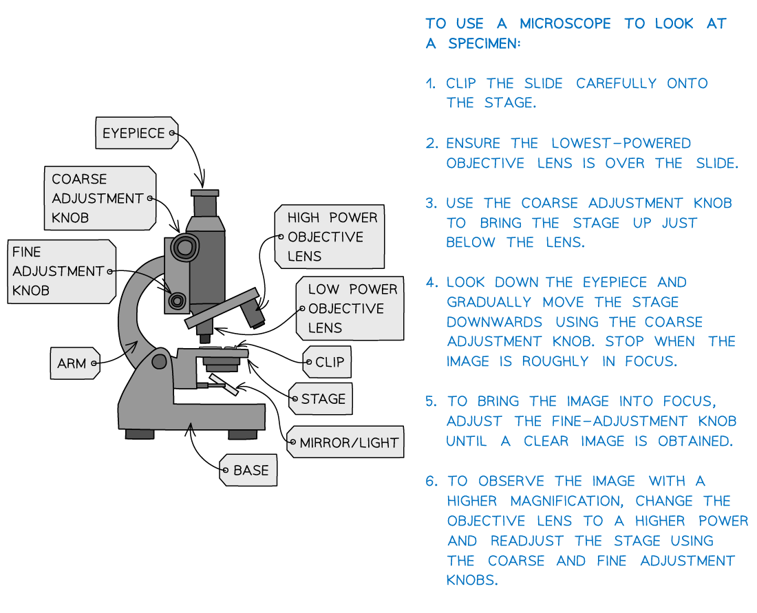
When using an optical microscope always start with the lowest power objective lens:
It is easier to find what you are looking for in the field of view
This helps to prevent damage to the lens or coverslip in case the stage has been raised too high
Preventing the dehydration of tissue:
The thin layers of material placed on slides can dry up rapidly
Adding a drop of water to the specimen (beneath the coverslip) can prevent the cells from being damaged by dehydration
Unclear or blurry images:
Switch to the lower power objective lens and try using the coarse focus to get a clearer image
Consider whether the specimen sample is thin enough for light to pass through to see the structures clearly
There could be cross-contamination with foreign cells or bodies
Viewing plant tissue
An ideal tissue is the onion epidermis (found between the layers of onions) because it forms a layer just one cell thick
Being a non-photosynthetic tissue, onion epidermis is not green as it does not contain any chloroplasts
Apparatus
The key components of an optical microscope you will need to use are:
The eyepiece lens
The objective lenses
The stage
The light source
The coarse and fine focus
Other apparatus used:
Forceps
Scissors
Scalpel
Coverslip
Slides
Pipette
Iodine solution
Viewing onion cells under the microscope diagram
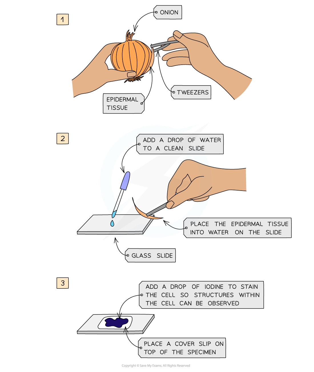
Care must be taken to avoid smudging the glass slide or trapping air bubbles under the coverslip
Viewing animal tissue
Human cheek cells are a good choice for examination under the light microscope because they are:
Plentiful
Easy to obtain safely
Can be obtained without an overly intrusive process
Relatively undifferentiated and so will display the main cell structures
Safety considerations
Do not perform the sampling on a person who has a cold, cough, throat infection etc.
To avoid spreading the infection to others
Concentrated methylene blue is toxic if ingested
Wear gloves and do NOT allow children to handle methylene blue solution or have access to the bottle of solution
Apparatus
Glass microscope slides
Cover slips
Paper towels or tissue
Staining solution
Methylene blue solution
0.5% to 1%
Dilute according to concentration of the stock solution
Plastic pipette or dropper
Sterile, individually packed cotton wool buds or swabs
Method
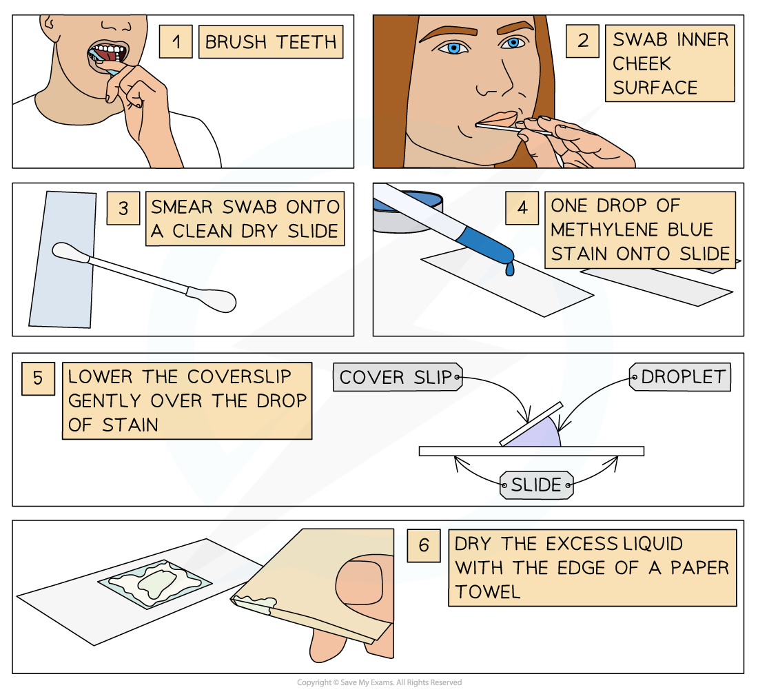
Brush teeth thoroughly with normal toothbrush and toothpaste
This removes bacteria from teeth so they don't obscure the view of the cheek cell
Take a clean, sterile cotton swab and gently scrape the inside cheek surface of the mouth for 5-10 seconds
Smear the cotton swab on the centre of the microscope slide for 2 to 3 seconds
Add a drop of methylene blue solution
Place a coverslip on top
Lay the coverslip down at one edge and then tilt it down flat
This reduces bubble formation under the coverslip
Absorb any excess solution by allowing a paper towel to touch one side of the coverslip.
Place the slide on the microscope, with 4 x or 10 objective in position and find a cell
Then view at higher magnification to reveal more detail
Methylene blue stains negatively charged molecules in the cell, including DNA and RNA
This causes the nucleus and mitochondria appear darker than their surroundings
The cells seen are squamous epithelial cells from the outer epithelial layer of the mouth
Viewing cheek cells under the microscope diagram
Parts of the cell that can be seen with a light microscope
Nucleus
Mitochondria
Cell membrane
Cytoplasm
Parts of the cell that cannot be seen with a light microscope
Ribosomes*
Endoplasmic reticulum*
Golgi*
Details of the nucleus, mitochondria and cell membrane
* these are parts of the cell that you don't need to know the names of, but they are included here as examples of very small structures within the cell