Development of the Zygote
Development of the Zygote
Development of the Zygote
After fertilisation in the oviduct, the zygote travels towards the uterus
This takes about 3 days, during which time the zygote will divide several times to form a ball of cells known as an embryo
In the uterus, the embryo embeds itself in the thick lining (implantation) and continues to grow and develop
The gestation period for humans is 9 months
Major development of organs takes place within the first 12 weeks, during which time the embryo gets nutrients from the mother by diffusion through the uterus lining
After this point, the organs are all in place, the placenta has formed and the embryo is now called a fetus
The remaining gestation time is used by the fetus to grow bigger in size
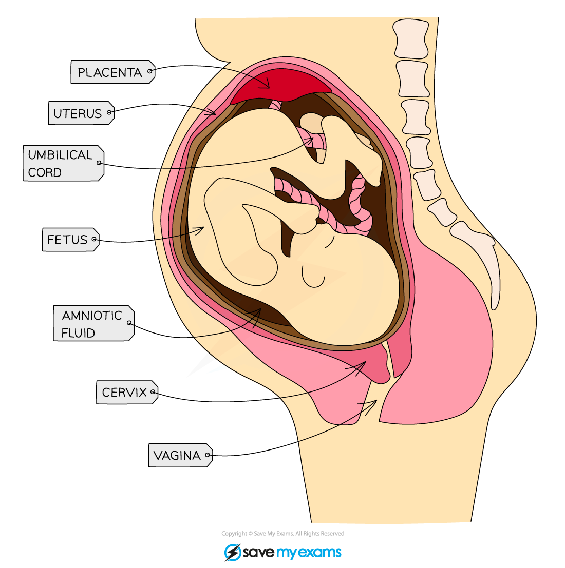
The foetus in the uterus
The fetus is surrounded by an amniotic sac which contains amniotic fluid (made from the mother’s blood plasma)
This protects the fetus during development by cushioning it from bumps to the mother’s abdomen
The umbilical cord joins the fetus’s blood supply to the placenta for exchange of nutrients and removal of waste products
Virus Transmission Across the Placenta
Virus Transmission Across the Placenta
During the gestation period the fetus develops and grows by gaining the glucose, amino acids, fats, water and oxygen it needs from the mother’s blood
The blood runs opposite each other, never mixing, in the placenta
The fetus’s blood connects to and from the placenta by the umbilical cord
The mother’s blood also absorbs the waste from the fetus’s blood in the placenta; substances like carbon dioxide and urea are removed from the fetus’s blood so that they do not build up to dangerous levels
Movement of all molecules across the placenta occurs by diffusion due to differences in concentration gradients
The placenta is adapted for this diffusion by having a large surface area and a thin wall for efficient diffusion
The placenta acts as a barrier to prevent toxins and pathogens from getting into the fetus’s blood
Not all toxin molecules or pathogenic organisms are stopped from passing through the placenta (this usually depends on the size of the molecule)
This is why pregnant women are advised not to smoke during pregnancy, as molecules like nicotine can pass across the placenta
Certain viruses are small enough to cross the placenta and reach the bloodstream of the fetus
Examples of such viruses include rubella and HIV
Once circulating in the blood of the fetus, these viruses can cause very serious health complications
Rubella may lead to problems such as deafness, heart disease and eye problems
HIV could be fatal to a foetus
The structure of the placenta diagram
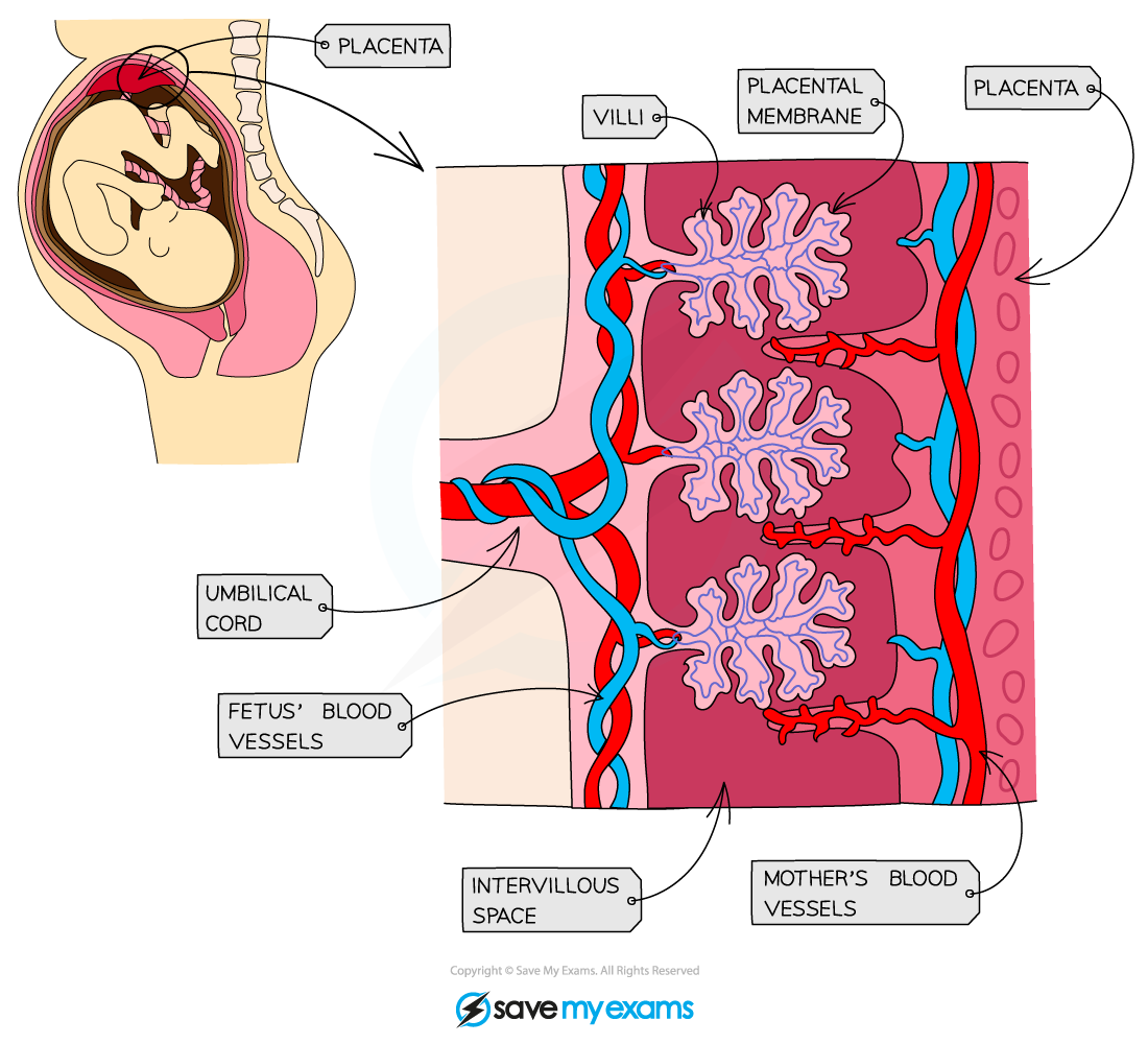
Certain molecules and viruses are small enough to cross the placental barrier and reach the bloodstream of the fetus